
 New Products
 Ordering
 Distributors
 Resources
 FAQs
FAQs Cart
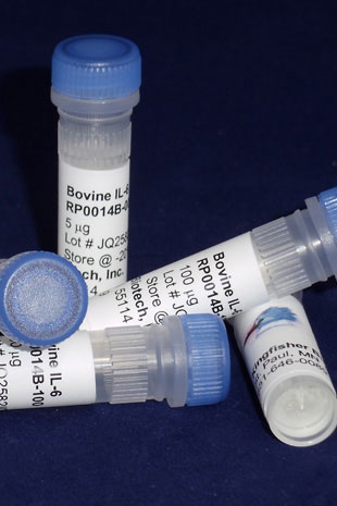
CartBovine IL-6 (Yeast-derived Recombinant Protein) - 25 micrograms
Bulk quantities of Bovine IL-6 protein are available.
Please contact us for pricing.
Bovine IL-6 Specifications
Molecular Weight (calculated) - 20.7kDa
Amino Acid Sequence - GPLGEDFKND TTPGRLLLTT PEKTEALIKR MVDKISAMRK EICEKNDECE SSKETLAENK LNLPKMEEKD GCFQSGFNQA ICLIRTTAGL LEYQIYLDYL QNEYEGNQEN VRDLRKNIRT LIQILKQKIA DLITTPATNT DLLEKMQSSN EWVKNAKIIL ILRNLENFLQ FSLRAIRMK (179)
Gene ID - 280826
Homology Across Species
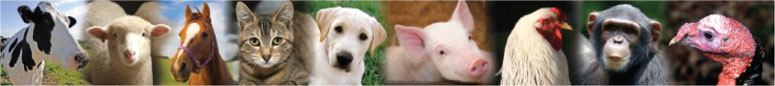
Bos taurus (cattle) IL-6 – 100%
Bos indicus (zebu) IL-6 – 100%
Bison bison (bison) IL-6 – 99%
Bos mutus (wild yak) IL-6 – 99%
Bubalus bubalis (water buffalo) IL-6 – 99%
Syncerus caffer (African buffalo) IL-6 – 98%
More - https://blast.ncbi.nlm.nih.gov/
Endotoxin - Naturally endotoxin-free
Applications
Cell Culture, ELISA Standard, Western Blot Control
IL-6 Background
Interleukin-6 (IL-6) is an interleukin that acts as both a pro-inflammatory and anti-inflammatory cytokine. It is secreted by T cells and macrophages to stimulate immune response to trauma, especially burns or other tissue damage leading to inflammation. IL-6 is also produced from muscle, and is elevated in response to muscle contraction. It is significantly elevated with exercise, and precedes the appearance of other cytokines in the circulation. Osteoblasts secrete IL-6 to stimulate osteoclast formation. Smooth muscle cells in the tunica media of many blood vessels also produce IL-6 as a pro-inflammatory cytokine. The role of IL-6 as an anti-inflammatory cytokine is mediated through its inhibitory effects on TNF-alpha and IL-1, and activation of IL-1ra and IL-10.
Alternate Names - IL6, BSF2, HGF, HSF, IFNB2, IL-6, BSF-2, CDF, IFN-beta-2, interleukin 6
Aerosol vaccination with Bacille CalmetteGuerin induces a trained innate immune phenotype in calves.
Guerra-Maupome M, Vang DX, McGill JL.
PLoS One. 2019 Feb 22;14(2):e0212751. doi: 10.1371/journal.pone.0212751. eCollection 2019.
Applications: Measurement of bovine TNF alpha, IL-1 beta, and IL-6 in culture supernatants by ELISA
Abstract
Mycobacterium bovis Bacillus Calmette-Guérin (BCG) is a live attenuated vaccine for use against tuberculosis (TB); however, it is known to reduce childhood mortality from infections other than TB. The unspecific protection induced by BCG vaccination has been associated with the induction of memory-like traits of the innate immune system identified as 'trained' immunity. In humans and mouse models, in vitro and in vivo BCG training leads to enhanced production of monocyte-derived proinflammatory cytokines in response to secondary unrelated bacterial and fungal pathogens. While BCG has been studied extensively for its ability to induce innate training in humans and mouse models, BCG's nonspecific protective effects have not been defined in agricultural species. Here, we show that in vitro BCG training induces a functional change in bovine monocytes, characterized by increased transcription of proinflammatory cytokines upon restimulation with the toll-like receptor agonists. Importantly, in vivo, aerosol BCG vaccination in young calves also induced a 'trained' phenotype in circulating peripheral blood mononuclear cells (PBMCs), that lead to a significantly enhanced TLR-induced proinflammatory cytokine response and changes in cellular metabolism compared to PBMCs from unvaccinated control calves. Similar to the long-term training effects of BCG reported in humans, our results suggest that in young calves, the effects of BCG induced innate training can last for at least 3 months in circulating immune populations. Interestingly, however, aerosol BCG vaccination did not 'train' the innate immune response at the mucosal level, as alveolar macrophages from aerosol BCG vaccinated calves did not mount an enhanced inflammatory response to secondary stimulation, compared to cells isolated from control calves. Together, our results suggest that, like mice and humans, the innate immune system of calves can be 'trained'; and that BCG vaccination could be used as an immunomodulatory strategy to reduce disease burden in juvenile food animals before the adaptive immune system has fully matured.
Interleukin-6 increases inner cell mass numbers in bovine embryos.
Wooldridge LK, Ealy AD.
BMC Dev Biol. 2019 Feb 1;19(1):2. doi: 10.1186/s12861-019-0182-z.
Applications: Stimulation of in vitro-produced bovine embryos
Abstract
BACKGROUND:
Work in other species suggests that interleukin-6 (IL6) promotes early embryo development. It was unclear whether IL6 serves as an embryokine in cultured bovine embryos. This work was undertaken to elucidate the role of IL6 during in vitro bovine embryo production.
RESULTS:
Transcripts for IL6 and its two cognate receptor subunits (IL6R, IL6ST) were confirmed in bovine embryos from the 1-cell to blastocyst stages. Supplementing 100 ng/ml recombinant bovine IL6 to in vitro-produced bovine embryos at day 1, 3 or 5 increased (P < 0.05) inner cell mass (ICM) cell number and the ICM:trophectoderm (TE) ratio but not TE cell number. No increase in ICM or TE cell number was observed after supplementation of 1 or 10 ng/ml IL6 beginning at either day 1 or 5. Sequential supplementation with 100 ng/ml IL6 at both day 1 and 5 (for a total of 200 ng/ml IL6) increased (P < 0.05) ICM cell number to a greater extent than supplementing IL6 at a single time period in one study but not a second study. Additionally, providing 200 ng/ml IL6 beginning at day 1 or 5 yielded no further increase on ICM cell numbers when compared to supplementing with 100 ng/ml IL6. IL6 treatment had no effect on cleavage or blastocyst formation in group culture. However, IL6 supplementation increased cleavage and day 8 blastocyst formation when bovine embryos were cultured individually.
CONCLUSIONS:
These results implicate IL6 as an embryokine that specifically increases ICM cell numbers in bovine embryos and facilitates bovine blastocyst development in embryos cultured individually.
Effect of mesenchymal precursor cells on the systemic inflammatory response and endothelial dysfunction in an ovine model of collagen-induced arthritis.
Dooley LM, Abdalmula A, Washington EA, Kaufman C, Tudor EM, Ghosh P, Itescu S, Kimpton WG, Bailey SR.
PLoS One. 2015 May 7;10(5):e0124144. doi: 10.1371/journal.pone.0124144. eCollection 2015.
Applications: ELISA standard
Abstract
BACKGROUND AND AIM:
Mesenchymal precursor cells (MPC) are reported to possess immunomodulatory properties that may prove beneficial in autoimmune and other inflammatory conditions. However, their mechanism of action is poorly understood. A collagen-induced arthritis model has been previously developed which demonstrates local joint inflammation and systemic inflammatory changes. These include not only increased levels of inflammatory markers, but also vascular endothelial cell dysfunction, characterised by reduced endothelium-dependent vasodilation. This study aimed to characterise the changes in systemic inflammatory markers and endothelial function following the intravenous administration of MPC, in the ovine model.
METHODS:
Arthritis was induced in sixteen adult sheep by administration of bovine type II collagen into the hock joint following initial sensitisation. After 24h, sheep were administered either 150 million allogeneic ovine MPCs intravenously, or saline only. Fibrinogen and serum amyloid-A were measured in plasma to assess systemic inflammation, along with pro-inflammatory and anti-inflammatory cytokines. Animals were necropsied two weeks following arthritis induction. Coronary and digital arterial segments were mounted in a Mulvaney-Halpern wire myograph. The relaxant response to endothelium-dependent and endothelium-independent vasodilators was used to assess endothelial dysfunction.
RESULTS AND CONCLUSION:
Arthritic sheep treated with MPC demonstrated a marked spike in plasma IL-10, 24h following MPC administration. They also showed significantly reduced plasma levels of the inflammatory markers, fibrinogen and serum amyloid A, and increased HDL. Coronary arteries from RA sheep treated with MPCs demonstrated a significantly greater maximal relaxation to bradykinin when compared to untreated RA sheep (253.6 ± 17.1% of pre-contracted tone vs. 182.3 ± 27.3% in controls), and digital arteries also demonstrated greater endothelium-dependent vasodilation. This study demonstrated that MPCs given intravenously are able to attenuate systemic inflammatory changes associated with a monoarthritis, including the development of endothelial dysfunction.
Adventitial fibroblasts induce a distinct proinflammatory/profibrotic macrophage phenotype in pulmonary hypertension
El Kasmi KC, Pugliese SC, Riddle SR, Poth JM, Anderson AL, Frid MG, Li M, Pullamsetti SS, Savai R, Nagel MA, Fini MA, Graham BB, Tuder RM, Friedman JE, Eltzschig HK, Sokol RJ, Stenmark KR.
J Immunol. 2014 Jul 15;193(2):597-609
Applications: Bovine IL-6 was quantified using an ELISA development kit. Bovine rIL-6 was used as a standard
Effects of interleukin-8 on estradiol and progesterone production by bovine granulosa cells from large follicles and progesterone production by luteinizing granulosa cells in culture.
Shimizu T, Kaji A, Murayama C, Magata F, Shirasuna K, Wakamiya K, Okuda K, Miyamoto A.
Cytokine. 2012 Jan;57(1):175-81.
Applications: Non-specific ELISA Standard
Abstract
Interleukin 8 (IL-8) is a chemoattractant involved in the recruitment and activation of neutrophils and is associated with the ovulate process. We examined the possible role of IL-8 in steroid production by bovine granulosa cells before and after ovulation. The concentration of IL-8 in the follicular fluid of estrogen-active dominant (EAD) and pre-ovulatory follicles (POF) was higher than that of small follicles (SF). CXCR1 mRNA expression was higher in the granulosa cells of EAD and POF than that of SF. In contrast, CXCR2 mRNA expression was lower in granulosa cells of EAD and POF than in SF. IL-8 inhibited estradiol (E2) production in follicle-stimulating hormone (FSH)-treated granulosa cells at 48 h of culture. IL-8 also suppressed CYP19A1 mRNA expression in FSH-treated granulosa cells. IL-8 stimulated progesterone (P4) production in luteinizing hormone (LH)-treated granulosa cells at 48 h of culture. Although IL-8 did not alter the expression of genes associated with P4 production, it induced StAR protein expression in LH-treated granulosa cells. The expression of CXCR1 mRNA in corpus luteum (CL) did not change during the luteal phase. In contrast, the expression of CXCR2 mRNA in middle CL was significantly higher than in early and regression CL during the luteal phase. In luteinizing granulosa cells, an in vitro model of granulosa cell luteinization, CXCR2 mRNA expression was downregulated, whereas CXCR1 mRNA expression was unchanged. IL-8 also stimulated P4 production in luteinizing granulosa cells. These data provide evidence that IL-8 functions not only as a chemokine, but also act as a regulator of steroid synthesis in granulosa cells to promote luteinization after ovulation.
Ordering Information & Terms and Conditions
We require a phone number and e-mail address for both the end user of the ordered product and your institution's Accounts Payable representative. This information is only used to help with technical and billing issues.
Via Phone
Please call us at 651-646-0089 between the hours of 8:30 a.m. and 5:30 p.m. CST Mon - Fri.
Via Fax
Orders can be faxed to us 24 hours a day at 651-646-0095.
Via E-mail
Please e-mail orders to orders@KingfisherBiotech.com.
Via Mail
Please mail your order to:
Sales Order Entry
Kingfisher Biotech, Inc.
1000 Westgate Drive
Suite 123
Saint Paul, MN 55114
USA
Product Warranty
Kingfisher Biotech brand products are warranted by Kingfisher Biotech, Inc. to meet stated product specifications and to conform to label descriptions when used, handled and stored according to instructions. Unless otherwise stated, this warranty is limited to one year from date of sale. Kingfisher Biotech’s sole liability for the product is limited to replacement of the product or refund of the purchase price. Kingfisher Biotech brand products are supplied for research applications. They are not intended for medicinal, diagnostic or therapeutic use. The products may not be resold, modified for resale or used to manufacture commercial products without prior written approval from Kingfisher Biotech.
Payment Terms
All prices are subject to change without notice. Payment terms are net thirty (30) days from receipt of invoice. A 1.5% service charge per month is added for accounts past due over 30 days. Prices quoted are U.S. Dollars. The purchaser assumes responsibility for any applicable tax. You will only be charged for products shipped. Products placed on back order will be charged when shipped. If you place an order and fail to fulfill the terms of payment, Kingfisher Biotech, Inc. may without prejudice to any other lawful remedy defer further shipments and/or cancel any order. You shall be liable to Kingfisher Biotech, Inc. for all costs and fees, including attorneys' fees, which Kingfisher Biotech, Inc. may reasonably incur in any actions to collect on your overdue account. Kingfisher Biotech, Inc. does not agree to, and is not bound by, any other terms or conditions such as terms in a purchase order that have not been expressly agreed to in writing signed by a duly authorized officer of Kingfisher Biotech, Inc.
Shipping
Shipping and handling costs are prepaid and added to the invoice. Shipping and handling costs will be charged only on the first shipment in situations where an order contains back ordered products. Kingfisher Biotech, Inc. reserves the right to select the packaging and shipping method for your order, which will ensure the stability of the product and also efficient tracing. Domestic orders will normally be shipped by overnight. Damage during shipment is covered by the warranty provided in these terms and conditions. For international orders, title to the goods passes in the United States when the goods are placed with the shipper. For all orders, the risk of loss of the goods passes when the goods are placed with the shipper.
Returns
Please call customer service before returning any products for refund, credit or replacement. NO returns will be accepted without prior written authorization. Returns are subject to a restocking fee of 20%.