

 New Products
 Ordering
 Distributors
 Resources
 FAQs
FAQs Cart
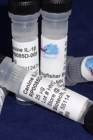
CartCanine IL-1 beta (IL-1F2) (Yeast-derived Recombinant Protein) - 25 micrograms
The IL-1 family of cytokines encompasses eleven proteins that each share a similar β-barrel structure and bind to Ig-like receptors. Several of the well characterized members of the IL-1-like cytokines play key roles in the development and regulation of inflammation. IL-1α (IL-1F1), IL-1β (IL-1F2), and IL-18 (IL-1F4) are well-known inflammatory cytokines active in the initiation of the inflammatory reaction and in driving Th1 and Th17 inflammatory responses. In contrast, IL-1 receptor antagonist (IL-1ra; IL-1F3) and IL-36 receptor antagonist (IL-36ra; IL-1F5) reduce inflammation by blocking the binding of the agonist receptor ligands. IL-33 (IL-1F11) is thought to function as an 'alarmin' released following cell necrosis to alerting the immune system to tissue damage or stress. The biological properties of IL-37 (IL-1F7) are mainly those of down-regulating inflammation.
Alternate Names - IL1B, IL-1, IL1-BETA, IL1F2, interleukin 1 beta, IL1beta
IL-1β Homology Across Species
Canis lupus familiaris (dog) IL-1β – 100%
Canis lupus dingo (dingo) IL-1β – 100%
Vulpes vulpes (red fox) IL-1β – 96%
More - https://blast.ncbi.nlm.nih.gov/
Elevated circulating Th2 but not group 2 innate lymphoid cell responses characterize canine atopic dermatitis.
Früh SP, Saikia M, Eule J, Mazulis CA, Miller JE, Cowulich JM, Oyesola OO, Webb LM, Peng SA, Cubitt RL, Danko CG, Miller WH, Tait Wojno ED.
Vet Immunol Immunopathol. 2020 Jan 24;221:110015. doi: 10.1016/j.vetimm.2020.110015. [Epub ahead of print]
Applications: In vitro culture of CD4+ T cells and CD25+ ILCs
ERK1/ATF-2 signaling axis contributes to interleukin-1β-induced MMP-3 expression in dermal fibroblasts.
Kitanaka N, Nakano R, Sakai M, Kitanaka T, Namba S, Konno T, Nakayama T, Sugiya H.
PLoS One. 2019 Sep 19;14(9):e0222869. doi: 10.1371/journal.pone.0222869. eCollection 2019.
Applications: Stimulation of canine dermal fibroblasts.
Abstract
Matrix metalloproteinases (MMPs) play a pivotal role in tissue remodeling by degrading the extracellular matrix (ECM) components. This mechanism is implicated in a variety of physiological and pathological cellular processes including wound healing. One of the key proteins involved in this process is the proinflammatory cytokine interleukin-1β (IL-1β, which induces the expression of MMP-3 mRNA and the secretion of MMP-3 protein by dermal fibroblasts. In this study, we first investigated the contribution of activating transcription factor 2 (ATF-2) to IL-1β-induced MMP-3 expression in dermal fibroblasts. Our results showed that in cells transfected with ATF-2 siRNA or treated with the ATF-2 inhibitor SBI-0087702, IL-1β-induced MMP-3 mRNA expression was reduced. We also demonstrated that IL-1β stimulates the phosphorylation of ATF-2. These observations suggest that ATF-2 plays an important role in IL-1β-induced MMP-3 expression. Next, we investigated the role of MAPK signaling in ATF-2 activation. In cells treated with the extracellular signal-regulated kinase (ERK) inhibitor FR180240, as well as in cells transfected with ERK1 and ERK2 siRNAs, IL-1β-induced MMP-3 mRNA expression was reduced. In addition, we showed that IL-1β induced the phosphorylation of ERK1/2. These observations suggest that ERK1 and ERK2 are involved in IL-1β-induced MMP-3 expression. However, ERK1 and ERK2 do seem to play different roles. While the ERK inhibitor FR180204 inhibited IL-1β-induced ATF-2 phosphorylation, only in cells transfected with ERK1 siRNA, but not ERK2 siRNA, IL-1β-induced ATF-2 phosphorylation was reduced. These findings suggest that the ERK1/ATF-2 signaling axis contributes to IL-1β-induced MMP-3 expression in dermal fibroblasts.
Interleukin-1β promotes interleulin-6 expression via ERK1/2 signaling pathway in canine dermal fibroblasts.
Kitanaka N, Nakano R, Sugiura K, Kitanaka T, Namba S, Konno T, Nakayama T, Sugiya H.
PLoS One. 2019 Jul 25;14(7):e0220262. doi: 10.1371/journal.pone.0220262. eCollection 2019.
Applications: Simulation of dermal fibroblasts in culture
Abstract
Interleukin-6 (IL-6) is a pleiotropic cytokine involved in the regulation of the immune response and inflammation. In this study, we investigated effect of the proinflammatory cytokine interleukin-1β (IL-1β) on IL-6 expression in canine dermal fibroblasts. IL-1β induced IL-6 mRNA expression and protein release in a time- and dose-dependent manner. When cells were treated with inhibitors of mitogen-activated protein kinases (MAPKs), the extracellular signal-regulated kinase (ERK) inhibitor FR180240 inhibited IL-1β-induced IL-6 mRNA expression, but not SP600125 or SKF86002, which are c-Jun N-terminal kinase (JNK) and p38 MAPK inhibitors, respectively. In cells treated with U0126, an inhibitor of MAPK/ERK kinase (MEK), which activates ERK, IL-1β-induced IL-6 mRNA expression was also inhibited. IL-1β stimulated ERK1/2 phosphorylation. In cells transfected with ERK1 and ERK2 isoform siRNAs, IL-1β-induced IL-6 mRNA expression was reduced. These observations suggest that IL-1β induces IL-6 expression via ERK1/2 signaling pathway in canine dermal fibroblasts.
Interleukin-1β expression is increased in the duodenum of dogs with idiopathic inflammatory bowel disease.
Hawes M, Riddle A, Kirk J, Jergens A, Allenspach K.
Vet Rec. 2018 Nov 3;183(17):536. doi: 10.1136/vr.104495. Epub 2018 May 9.
Applications: Measurement of canine IL-1 beta in tissue homogenates supernatant in dogs with IBD by ELISA.
Cytokine and Growth Factor Concentrations in Canine Autologous Conditioned Serum.
Sawyere DM, Lanz OI, Dahlgren LA, Barry SL, Nichols AC, Werre SR.
Vet Surg. 2016 Jul;45(5):582-6. doi: 10.1111/vsu.12506.
Applications: Measurement of canine IL-1RA, IL-1 beta, and TNF alpha in serum and plasma by ELISA
Th17 Pathway As a Target for Multipotent Stromal Cell Therapy in Dogs: Implications for Translational Research.
Kol A, Walker NJ, Nordstrom M, Borjesson DL.
PLoS One. 2016 Feb 12;11(2):e0148568. doi: 10.1371/journal.pone.0148568. eCollection 2016.
Applications: Canine IL-6 and IL-1 beta proteins were use to stimulate T cells in culture.
Abstract
Detrimental Th17 driven inflammatory and autoimmune disease such as Crohn's disease, graft versus host disease and multiple sclerosis remain a significant cause of morbidity and mortality worldwide. Multipotent stromal/stem cell (MSC) inhibit Th17 polarization and activation in vitro and in rodent models. As such, MSC based therapeutic approaches are being investigated as novel therapeutic approaches to treat Th17 driven diseases in humans. The significance of naturally occurring diseases in dogs is increasingly recognized as a realistic platform to conduct pre-clinical testing of novel therapeutics. Full characterization of Th17 cells in dogs has not been completed. We have developed and validated a flow-cytometric method to detect Th17 cells in canine blood. We further demonstrate that Th17 and other IL17 producing cells are present in tissues of dogs with naturally occurring chronic inflammatory diseases. Finally, we have determined the kinetics of a canine specific Th17 polarization in vitro and demonstrate that canine MSC inhibit Th17 polarization in vitro, in a PGE2 independent mechanism. Our findings provide fundamental research tools and suggest that naturally occurring diseases in dogs, such as inflammatory bowel disease, may be harnessed to translate novel MSC based therapeutic strategies that target the Th17 pathway.
Interleukin-1β, tumour necrosis factor-α and lipopolysaccharide induce C-type natriuretic peptide from canine aortic endothelial cells.
Osterbur K, Yu DH, Declue AE.
Res Vet Sci. 2012 Nov 8. doi:pii: S0034-5288(12)00305-0. 10.1016/j.rvsc.2012.10.002.
Applications: Stimulation of canine aortic endothelial cells
Ordering Information & Terms and Conditions
We require a phone number and e-mail address for both the end user of the ordered product and your institution's Accounts Payable representative. This information is only used to help with technical and billing issues.
Via Phone
Please call us at 651-646-0089 between the hours of 8:30 a.m. and 5:30 p.m. CST Mon - Fri.
Via Fax
Orders can be faxed to us 24 hours a day at 651-646-0095.
Via E-mail
Please e-mail orders to orders@KingfisherBiotech.com.
Via Mail
Please mail your order to:
Sales Order Entry
Kingfisher Biotech, Inc.
1000 Westgate Drive
Suite 123
Saint Paul, MN 55114
USA
Product Warranty
Kingfisher Biotech brand products are warranted by Kingfisher Biotech, Inc. to meet stated product specifications and to conform to label descriptions when used, handled and stored according to instructions. Unless otherwise stated, this warranty is limited to one year from date of sale. Kingfisher Biotech’s sole liability for the product is limited to replacement of the product or refund of the purchase price. Kingfisher Biotech brand products are supplied for research applications. They are not intended for medicinal, diagnostic or therapeutic use. The products may not be resold, modified for resale or used to manufacture commercial products without prior written approval from Kingfisher Biotech.
Payment Terms
All prices are subject to change without notice. Payment terms are net thirty (30) days from receipt of invoice. A 1.5% service charge per month is added for accounts past due over 30 days. Prices quoted are U.S. Dollars. The purchaser assumes responsibility for any applicable tax. You will only be charged for products shipped. Products placed on back order will be charged when shipped. If you place an order and fail to fulfill the terms of payment, Kingfisher Biotech, Inc. may without prejudice to any other lawful remedy defer further shipments and/or cancel any order. You shall be liable to Kingfisher Biotech, Inc. for all costs and fees, including attorneys' fees, which Kingfisher Biotech, Inc. may reasonably incur in any actions to collect on your overdue account. Kingfisher Biotech, Inc. does not agree to, and is not bound by, any other terms or conditions such as terms in a purchase order that have not been expressly agreed to in writing signed by a duly authorized officer of Kingfisher Biotech, Inc.
Shipping
Shipping and handling costs are prepaid and added to the invoice. Shipping and handling costs will be charged only on the first shipment in situations where an order contains back ordered products. Kingfisher Biotech, Inc. reserves the right to select the packaging and shipping method for your order, which will ensure the stability of the product and also efficient tracing. Domestic orders will normally be shipped by overnight. Damage during shipment is covered by the warranty provided in these terms and conditions. For international orders, title to the goods passes in the United States when the goods are placed with the shipper. For all orders, the risk of loss of the goods passes when the goods are placed with the shipper.
Returns
Please call customer service before returning any products for refund, credit or replacement. NO returns will be accepted without prior written authorization. Returns are subject to a restocking fee of 20%.