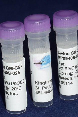
Swine GM-CSF (Yeast-derived Recombinant Protein) - 5 micrograms
Bulk quantities of proteins are available. Please contact us for bulk pricing.
Granulocyte-macrophage colony-stimulating factor (GM-CSF) is a protein secreted by macrophages, T cells, mast cells, NK cells, endothelial cells and fibroblasts. GM-CSF stimulates stem cells to produce granulocytes (neutrophils, eosinophils, and basophils) and monocytes. Granulocyte macrophage–colony stimulating factor (GM-CSF) is secreted in response to inflammatory stimuli such as LPS, IL-1, and TNF-α by a variety of different cells, including endothelium, fibroblasts, muscle cells, and macrophages, and by activated T cells. GM-CSF is glycosylated in its mature form.
Alternate Names - CSF2, GMCSF, colony stimulating factor 2, CSF
Homology Across Species
Sus scrofa (pig) GM-CSF – 100%
More - https://blast.ncbi.nlm.nih.gov/

A Nanoparticle-Poly(I:C) Combination Adjuvant Enhances the Breadth of the Immune Response to Inactivated Influenza Virus Vaccine in Pigs.
Renu S, Feliciano-Ruiz N, Lu F, Ghimire S, Han Y, Schrock J, Dhakal S, Patil V, Krakowka S, HogenEsch H, Renukaradhya GJ.
Vaccines (Basel). 2020 May 18;8(2):E229. doi: 10.3390/vaccines8020229.
Applications: IL-4 and GM-CSF were used to generate dendritic cells from porcine monocytes in culture.
Abstract
Intranasal vaccination elicits secretory IgA (SIgA) antibodies in the airways, which is required for cross-protection against influenza. To enhance the breadth of immunity induced by a killed swine influenza virus antigen (KAg) or conserved T cell and B cell peptides, we adsorbed the antigens together with the TLR3 agonist poly(I:C) electrostatically onto cationic alpha-D-glucan nanoparticles (Nano-11) resulting in Nano-11-KAg-poly(I:C) and Nano-11-peptides-poly(I:C) vaccines. In vitro, increased TNF-α and IL-1ß cytokine mRNA expression was observed in Nano-11-KAg-poly(I:C)-treated porcine monocyte-derived dendritic cells. Nano-11-KAg-poly(I:C), but not Nano-11-peptides-poly(I:C), delivered intranasally in pigs induced high levels of cross-reactive virus-specific SIgA antibodies secretion in the nasal passage and lungs compared to a multivalent commercial influenza virus vaccine administered intramuscularly. The commercial and Nano-11-KAg-poly(I:C) vaccinations increased the frequency of IFNγ secreting T cells. The poly(I:C) adjuvanted Nano-11-based vaccines increased various cytokine mRNA expressions in lymph nodes compared to the commercial vaccine. In addition, Nano-11-KAg-poly(I:C) vaccine elicited high levels of virus neutralizing antibodies in bronchoalveolar lavage fluid. Microscopic lung lesions and challenge virus load were partially reduced in poly(I:C) adjuvanted Nano-11 and commercial influenza vaccinates. In conclusion, compared to our earlier study with Nano-11-KAg vaccine, addition of poly(I:C) to the formulation improved cross-protective antibody and cytokine response.
Generation of human endothelium in pig embryos deficient in ETV2.
Das S, Koyano-Nakagawa N, Gafni O, Maeng G, Singh BN, Rasmussen T, Pan X, Choi KD, Mickelson D, Gong W, Pota P, Weaver CV, Kren S, Hanna JH, Yannopoulos D, Garry MG, Garry DJ.
Nat Biotechnol. 2020 Mar;38(3):297-302. doi: 10.1038/s41587-019-0373-y. Epub 2020 Feb 24.
Applications: The proteins were used in a hematopoietic assay which used cells from embryoid bodies.
Abstract
The scarcity of donor organs may be addressed in the future by using pigs to grow humanized organs with lower potential for immunological rejection after transplantation in humans. Previous studies have demonstrated that interspecies complementation of rodent blastocysts lacking a developmental regulatory gene can generate xenogeneic pancreas and kidney1,2. However, such organs contain host endothelium, a source of immune rejection. We used gene editing and somatic cell nuclear transfer to engineer porcine embryos deficient in ETV2, a master regulator of hematoendothelial lineages3-7. ETV2-null pig embryos lacked hematoendothelial lineages and were embryonic lethal. Blastocyst complementation with wild-type porcine blastomeres generated viable chimeric embryos whose hematoendothelial cells were entirely donor-derived. ETV2-null blastocysts were injected with human induced pluripotent stem cells (hiPSCs) or hiPSCs overexpressing the antiapoptotic factor BCL2, transferred to synchronized gilts and analyzed between embryonic day 17 and embryonic day 18. In these embryos, all endothelial cells were of human origin.
Ordering Information & Terms and Conditions
We require a phone number and e-mail address for both the end user of the ordered product and your institution's Accounts Payable representative. This information is only used to help with technical and billing issues.
Via Phone
Please call us at 651-646-0089 between the hours of 8:30 a.m. and 5:30 p.m. CST Mon - Fri.
Via Fax
Orders can be faxed to us 24 hours a day at 651-646-0095.
Via E-mail
Please e-mail orders to orders@KingfisherBiotech.com.
Via Mail
Please mail your order to:
Sales Order Entry
Kingfisher Biotech, Inc.
1000 Westgate Drive
Suite 123
Saint Paul, MN 55114
USA
Product Warranty
Kingfisher Biotech brand products are warranted by Kingfisher Biotech, Inc. to meet stated product specifications and to conform to label descriptions when used, handled and stored according to instructions. Unless otherwise stated, this warranty is limited to one year from date of sale. Kingfisher Biotech’s sole liability for the product is limited to replacement of the product or refund of the purchase price. Kingfisher Biotech brand products are supplied for research applications. They are not intended for medicinal, diagnostic or therapeutic use. The products may not be resold, modified for resale or used to manufacture commercial products without prior written approval from Kingfisher Biotech.
Payment Terms
All prices are subject to change without notice. Payment terms are net thirty (30) days from receipt of invoice. A 1.5% service charge per month is added for accounts past due over 30 days. Prices quoted are U.S. Dollars. The purchaser assumes responsibility for any applicable tax. You will only be charged for products shipped. Products placed on back order will be charged when shipped. If you place an order and fail to fulfill the terms of payment, Kingfisher Biotech, Inc. may without prejudice to any other lawful remedy defer further shipments and/or cancel any order. You shall be liable to Kingfisher Biotech, Inc. for all costs and fees, including attorneys' fees, which Kingfisher Biotech, Inc. may reasonably incur in any actions to collect on your overdue account. Kingfisher Biotech, Inc. does not agree to, and is not bound by, any other terms or conditions such as terms in a purchase order that have not been expressly agreed to in writing signed by a duly authorized officer of Kingfisher Biotech, Inc.
Shipping
Shipping and handling costs are prepaid and added to the invoice. Shipping and handling costs will be charged only on the first shipment in situations where an order contains back ordered products. Kingfisher Biotech, Inc. reserves the right to select the packaging and shipping method for your order, which will ensure the stability of the product and also efficient tracing. Domestic orders will normally be shipped by overnight. Damage during shipment is covered by the warranty provided in these terms and conditions. For international orders, title to the goods passes in the United States when the goods are placed with the shipper. For all orders, the risk of loss of the goods passes when the goods are placed with the shipper.
Returns
Please call customer service before returning any products for refund, credit or replacement. NO returns will be accepted without prior written authorization. Returns are subject to a restocking fee of 20%.


 New Products
 Ordering
 Distributors
 Resources
 FAQs
FAQs Cart
Cart