Canine TNF alpha Polyclonal Antibody
Tumor necrosis factor alpha (TNFSF2) is a member of the TNF Superfamily. It is produced chiefly by activated macrophages, but it is produced also by a broad variety of cell types including lymphoid cells, mast cells, endothelial cells, cardiac myocytes, adipose tissue, fibroblasts, and neuronal tissue. The primary role of TNF alpha is in the regulation of immune cells. TNF alpha, being an endogenous pyrogen, is able to induce fever, to induce apoptotic cell death, to induce sepsis (through IL-1 & IL-6 production), to induce cachexia, induce inflammation, and to inhibit tumorigenesis and viral replication.
Reactivity - ELISA
Bovine TNFα - None
Canine TNFα - Strong
Caprine TNFα - None
Cynomolgus Monkey TNFα - Strong
Dolphin TNFα - None
Equine TNFα - Weak
Feline TNFα - Moderate
Guinea Pig TNFα - None
Human TNFα - Strong
Mouse TNFα - None
Ovine TNFα - None
Rabbit TNFα - Moderate
Swine TNFα - None
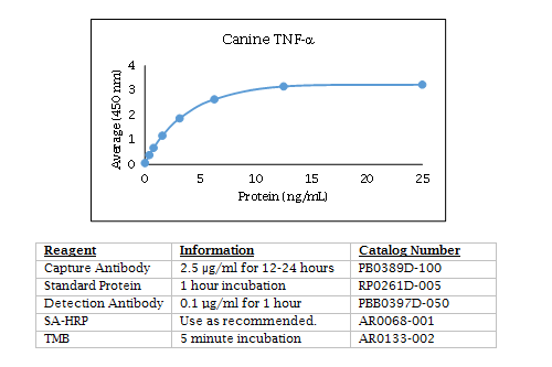
Canine TNFα ELISA Data

Blood and tissue biomarker analysis in dogs with osteosarcoma treated with palliative radiation and intra-tumoral autologous natural killer cell transfer.
Judge SJ, Yanagisawa M, Sturgill IR, Bateni SB, Gingrich AA, Foltz JA, Lee DA, Modiano JF, Monjazeb AM, Culp WTN, Rebhun RB, Murphy WJ, Kent MS, Canter RJ.
PLoS One. 2020 Feb 21;15(2):e0224775. doi: 10.1371/journal.pone.0224775. eCollection 2020.
Applications: Measurement of canine IL-2, and TNF alpha levels in serum by ELISA
Abstract
We have previously reported radiation-induced sensitization of canine osteosarcoma (OSA) to natural killer (NK) therapy, including results from a first-in-dog clinical trial. Here, we report correlative analyses of blood and tissue specimens for signals of immune activation in trial subjects. Among 10 dogs treated with palliative radiotherapy (RT) and intra-tumoral adoptive NK transfer, we performed ELISA on serum cytokines, flow cytometry for immune phenotype of PBMCs, and PCR on tumor tissue for immune-related gene expression. We then queried The Cancer Genome Atlas (TCGA) to evaluate the association of cytotoxic/immune-related gene expression with human sarcoma survival. Updated survival analysis revealed five 6-month survivors, including one dog who lived 17.9 months. Using feeder line co-culture for NK expansion, we observed maximal activation of dog NK cells on day 17-19 post isolation with near 100% expression of granzyme B and NKp46 and high cytotoxic function in the injected NK product. Among dogs on trial, we observed a trend for higher baseline serum IL-6 to predict worse lung metastasis-free and overall survival (P = 0.08). PCR analysis revealed low absolute gene expression of CD3, CD8, and NKG2D in untreated OSA. Among treated dogs, there was marked heterogeneity in the expression of immune-related genes pre- and post-treatment, but increases in CD3 and CD8 gene expression were higher among dogs that lived > 6 months compared to those who did not. Analysis of the TCGA confirmed significant differences in survival among human sarcoma patients with high and low expression of genes associated with greater immune activation and cytotoxicity (CD3e, CD8a, IFN-γ, perforin, and CD122/IL-2 receptor beta). Updated results from a first-in-dog clinical trial of palliative RT and autologous NK cell immunotherapy for OSA illustrate the translational relevance of companion dogs for novel cancer therapies. Similar to human studies, analyses of immune markers from canine serum, PBMCs, and tumor tissue are feasible and provide insight into potential biomarkers of response and resistance.
Cytokine and Growth Factor Concentrations in Canine Autologous Conditioned Serum.
Sawyere DM, Lanz OI, Dahlgren LA, Barry SL, Nichols AC, Werre SR.
Vet Surg. 2016 Jul;45(5):582-6. doi: 10.1111/vsu.12506.
Applications: Measurement of canine IL-1RA, IL-1 beta, and TNF alpha in serum and plasma by ELISA
Ordering Information & Terms and Conditions
We require a phone number and e-mail address for both the end user of the ordered product and your institution's Accounts Payable representative. This information is only used to help with technical and billing issues.
Via Phone
Please call us at 651-646-0089 between the hours of 8:30 a.m. and 5:30 p.m. CST Mon - Fri.
Via Fax
Orders can be faxed to us 24 hours a day at 651-646-0095.
Via E-mail
Please e-mail orders to orders@KingfisherBiotech.com.
Via Mail
Please mail your order to:
Sales Order Entry
Kingfisher Biotech, Inc.
1000 Westgate Drive
Suite 123
Saint Paul, MN 55114
USA
Product Warranty
Kingfisher Biotech brand products are warranted by Kingfisher Biotech, Inc. to meet stated product specifications and to conform to label descriptions when used, handled and stored according to instructions. Unless otherwise stated, this warranty is limited to one year from date of sale. Kingfisher Biotech’s sole liability for the product is limited to replacement of the product or refund of the purchase price. Kingfisher Biotech brand products are supplied for research applications. They are not intended for medicinal, diagnostic or therapeutic use. The products may not be resold, modified for resale or used to manufacture commercial products without prior written approval from Kingfisher Biotech.
Payment Terms
All prices are subject to change without notice. Payment terms are net thirty (30) days from receipt of invoice. A 1.5% service charge per month is added for accounts past due over 30 days. Prices quoted are U.S. Dollars. The purchaser assumes responsibility for any applicable tax. You will only be charged for products shipped. Products placed on back order will be charged when shipped. If you place an order and fail to fulfill the terms of payment, Kingfisher Biotech, Inc. may without prejudice to any other lawful remedy defer further shipments and/or cancel any order. You shall be liable to Kingfisher Biotech, Inc. for all costs and fees, including attorneys' fees, which Kingfisher Biotech, Inc. may reasonably incur in any actions to collect on your overdue account. Kingfisher Biotech, Inc. does not agree to, and is not bound by, any other terms or conditions such as terms in a purchase order that have not been expressly agreed to in writing signed by a duly authorized officer of Kingfisher Biotech, Inc.
Shipping
Shipping and handling costs are prepaid and added to the invoice. Shipping and handling costs will be charged only on the first shipment in situations where an order contains back ordered products. Kingfisher Biotech, Inc. reserves the right to select the packaging and shipping method for your order, which will ensure the stability of the product and also efficient tracing. Domestic orders will normally be shipped by overnight. Damage during shipment is covered by the warranty provided in these terms and conditions. For international orders, title to the goods passes in the United States when the goods are placed with the shipper. For all orders, the risk of loss of the goods passes when the goods are placed with the shipper.
Returns
Please call customer service before returning any products for refund, credit or replacement. NO returns will be accepted without prior written authorization. Returns are subject to a restocking fee of 20%.


 New Products
 Ordering
 Distributors
 Resources
 FAQs
FAQs Cart
Cart